Sutureless Plastic Gastroschisis Repair in Perspective of a Developing Country: A Case Report

  • Syeda Namayah Fatima Hussain Liaquat National Hospital & Medical College
  • Muhammad Arshad, Prof.
  • Manal Nasir
Keywords: Gastroschisis, Plastic Sutureless Closure, Neonate

Abstract

Background: Gastroschisis is congenital abdominal wall defect in neonates which needs to be addressed immediately after birth. Various techniques for closure of the defect have been described in literature.

Case Report: We describe a sutureless closure of abdominal wall defect in a 1-day old newborn with gastroschisis. 

Conclusion: Plastic sutureless closure could be the preferred technique for managing gastroschisis in resource constrained countries.

Downloads

Download data is not yet available.

References

Jones AM, Isenburg J, Salemi JL, Arnold KE, Mai CT, Aggarwal D, et al. Increasing prevalence of gastroschisis--14 States, 1995-2012. MMWR Morb Mortal Wkly Rep. 2016; 65:23-6.

Wittekindt B, Schloesser R, Doberschuetz N, Salzmann-Manrique E, Grossmann J, Misselwitz B, et al. Epidemiology and outcome of major congenital malformations in a large german county. Eur J Pediatr Surg. 2019; 29:282-9.

Sandler A, Lawrence J, Meehan J, Phearman L, Soper R. A plastic sutureless abdominal wall closure in gastroschisis. J Pediatr Surg. 2004; 39:738-41.

Wright NJ, Langer M, Norman IC, Akhbari M, Wafford QE, Ade-Ajayi N, et al. Improving outcomes for neonates with gastroschisis in low-income and middle-income countries: a systematic review protocol. BMJ Paediatr Open. 2018; 2:e000392.

Kidd JN, Levy MS, Wagner CW. Staged reduction of gastroschisis: a simple method. Pediatr Surg Int. 2001; 17:242-4.

Bianchi A, Dickson AP. Elective delayed reduction and no anesthesia: 'minimal intervention management' for gastrochisis. J Pediatr Surg. 1998; 33:1338-40.

Lee SC, Jung SE, Kim WK. Silo formation without suturing in gastroschisis: use of Steridrape for delayed repair. J Pediatr Surg. 1997; 32:66-8.

Miyake H, Seo S, O'Connell JS, Janssen Lok M, Pierro A. Safety and usefulness of plastic closure in infants with gastroschisis: a systematic review and meta-analysis. Pediatr Surg Int. 2019; 35:107-16.

Bruzoni M, Jaramillo JD, Dunlap JL, Abrajano C, Stack SW, Hintz SR, et al. Sutureless vs sutured gastroschisis closure: A prospective randomized controlled trial. J Am Coll Surg. 2017; 224:1091-6 e1.
Published
2019-11-10
How to Cite
1.
Hussain SNF, Arshad M, Nasir M. Sutureless Plastic Gastroschisis Repair in Perspective of a Developing Country: A Case Report. APSP J Case Rep [Internet]. 10Nov.2019 [cited 11Oct.2024];10:4. Available from: http://www.apspjcaserep.com/ojs1/index.php/ajcr/article/view/92
Section
Case Reports and Letters

INTRODUCTION

Gastroschisis (incidence of 2-5 cases per 10,000 livebirths) is anterior abdominal wall defect occurring usually on the right side of umbilicus, leading to evisceration of abdominal contents.[1,2] The conventional methods used are; primary fascial repair. or staged silo reduction followed by a sutured closure of the abdominal wall. Plastic sutureless closure, as introduced by Sandler et al [3], used a combination of Tegaderm dressing and remnants of umbilical cord to cover the defect. Despite having a similar outcome, the plastic sutureless closure is considerable cost effective. We report a case of gastroschisis in a neonate managed by the plastic sutureless technique. To the best of our knowledge, this is the only documentation of this technique from Pakistan.

CASE REPORT

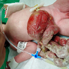
A one-day old full term baby girl, born by SVD and weighing 2.4 kilogram, presented with eviscerated bowel loops from abdominal wall. On examination, the stomach, small and large bowels were eviscerated through an abdominal wall defect on right side of umbilical cord (Figure 1). The organs had been covered in cotton wool and were densly adherent to each other. Laboratory work-up was normal, except a blood culture positive for Acinetobacter. Antibiotic therapy with vancomycin, meropenem and colomycin was started along with fluid resuscitation. After rapid optimization, the baby underwent surgery. After cleaning and washing, the eviscerated gut loops were reduced gently into the peritoneal cavity while noting intragastric pressure. Owing to difficulty in reduction, the defect was extended slightly. The defect was then covered by the umbilical cord cut according to the dimensions of the opening. Tegaderm dressing was then applied over the defect for reinforcement (Figure 2).

Figure 1: Neonate born with gastroschisis bowel before reduction. Stomach and edematous large bowel can be noted with stuck cotton used to clean it at home.
Figure 2: Post-operative picture showing closure of defect, done by primary reduction and tailored umbilical cord filling, covered by Tegaderm dressing.

After surgery, the baby was kept in NICU on ventilator and monitored. Repeat blood culture done post-operatively was negative. She was started on peripheral parenteral nutrition the next day and switched to oral feeding, gradually starting from the 6th post-operative day. The Tegaderm dressing was changed and the wound was washed with normal saline every 72 hours. On 4th post-operative day, purulent fluid started oozing from the wound. Peritoneal fluid was then sent for culture turned positive for Candida Tropicalis and Acinetobacter (Multi Drugs Resistant). The patient was treated with colomycin and fluconazole for the respective organisms. She was discharged on the 10th postoperative day in good condition. On follow up, the child is now 6 months old and doing well (Figure 3). She has developed a small umbilical hernia which is being managed expectantly.

Figure 3: Follow-up picture of the patient with a healed incision and the umbilical cord sloughed off.

DISCUSSION

Gastroschisis is a common abdominal wall defect in the neonates and its management is aimed at returning the eviscerated contents back into the abdominal cavity followed by the closure of the defect. Despite being a manageable condition, it has a high mortality rate with contrasting statistics; 75% to 100% in low middle income countries and less than 4% in high income countries, establishing the essential role of surgical management in survival.[4]

A number of surgical techniques have been described in literature which include primary closure, staged closure and plastic sutureless closure.[5-7] The plastic sutureless method involves reduction of abdominal viscera into the cavity followed by an umbilical remnant covering reinforced by Tegaderm dressings. This technique is either used as primary or secondary after silo placement, however in our case primary intervention after bowel reduction was undertaken.

A meta-analysis by Miyaki et al, showed plastic closure as a cost effective technique as it decreases ventilator time and hospital stay.[8] Since sutureless closure can also be employed as a bedside technique, it avoids the added cost of operation theatres and general anesthesia. Whereas, staged silo reduction technique required longer ventilatory support and hospital stay. Both the monetary cost and risk of post-operative infection are deemed much lower in the sutureless closure as the procedure involves the use of umbilical cord which is an autologous tissue. This technique also has less risk of infections[9] Similarly, we were able to wash the wound with normal saline at the time of dressing change which helped cleaning the pus thus treating the infection quickly. Moreover, the cosmetic outcome is quite acceptable due to cicatrization and secondary healing leading to a centrally positioned umbilicus.[3]

In conclusion, plastic sutureless technique is a cost effective treatment modality especially for resource constrained countries. The technique also has added benefits of less chances of infections and short hospital stay.

Conflicts of interest. None declared

Source of Support: Nil

Author contributions: All authors contributed equally in concept, drafting, literature review, and approval of final manuscript.

Consent Statement: Authors declared that they have informed written consent for publication of this case report including clinical material (photographs/video), and every effort will be made to conceal the identity of the patient although not fully guaranteed.

  1. (). . Jones AM, Isenburg J, Salemi JL, Arnold KE, Mai CT, Aggarwal D, et al. Increasing prevalence of gastroschisis--14 States, 1995-2012. MMWR Morb Mortal Wkly Rep. 2016; 65:23-6.
  2. (). . Wittekindt B, Schloesser R, Doberschuetz N, Salzmann-Manrique E, Grossmann J, Misselwitz B, et al. Epidemiology and outcome of major congenital malformations in a large german county. Eur J Pediatr Surg. 2019; 29:282-9.
  3. (). . Sandler A, Lawrence J, Meehan J, Phearman L, Soper R. A plastic sutureless abdominal wall closure in gastroschisis. J Pediatr Surg. 2004; 39:738-41.
  4. (). . Wright NJ, Langer M, Norman IC, Akhbari M, Wafford QE, Ade-Ajayi N, et al. Improving outcomes for neonates with gastroschisis in low-income and middle-income countries: a systematic review protocol. BMJ Paediatr Open. 2018; 2:e000392.
  5. (). . Kidd JN, Levy MS, Wagner CW. Staged reduction of gastroschisis: a simple method. Pediatr Surg Int. 2001; 17:242-4.
  6. (). . Bianchi A, Dickson AP. Elective delayed reduction and no anesthesia: 'minimal intervention management' for gastrochisis. J Pediatr Surg. 1998; 33:1338-40.
  7. (). . Lee SC, Jung SE, Kim WK. Silo formation without suturing in gastroschisis: use of Steridrape for delayed repair. J Pediatr Surg. 1997; 32:66-8.
  8. (). . Miyake H, Seo S, O'Connell JS, Janssen Lok M, Pierro A. Safety and usefulness of plastic closure in infants with gastroschisis: a systematic review and meta-analysis. Pediatr Surg Int. 2019; 35:107-16.
  9. (). . Bruzoni M, Jaramillo JD, Dunlap JL, Abrajano C, Stack SW, Hintz SR, et al. Sutureless vs sutured gastroschisis closure: A prospective randomized controlled trial. J Am Coll Surg. 2017; 224:1091-6 e1.