
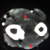
Periosteal Desmoplastic Fibroma of Radius: A Rare Bone Tumor
Downloads
References
Nedopil A, Raab P, Rudert M. Desmoplastic fibroma: a case report with three years of clinical and radiographic observation and review of the literature. Open Orthopaed J. 2013; 8:40.
Schneider M, Zimmermann AC, Depprich RA, Kübler NR, Engers R, Naujoks CD, et al. Desmoplastic fibroma of the mandible-review of the literature and presentation of a rare case. Head Face Med. 2009; 5:25.
Evans S, Ramasamy A, Jeys L, Grimer R. Desmoplastic fibroma of bone: A rare bone tumour. J Bone Oncol. 2014; 3: 77–9.
Crim JR, Gold RH, Mirra JM, Eckardt JJ, Bassett LW. Desmoplastic fibroma of bone: radiographic analysis. Radiology. 1989; 172: 827-32.
Jamsheidi K, Bagherifard A, Mirzaei A. Desmoplastic fibroma versus soft-tissue desmoid tumour of forearm: a case series of diagnosis, surgical approach, and outcome. Eur J Hand Surg. 2017; 42: 952-8.
Nuyttens JJ, Rust PF, Thomas Jr CR, Turrisi III AT. Surgery versus radiation therapy for patients with aggressive fibromatosis or desmoid tumors: a comparative review of 22 articles. Cancer. 2000; 88:1517-23.
RUI J, Guan W, GU y, Lao J. Treatment and functional result of Desmoplastic fibroma with repeated recurrences in the forearm: A case report. Onco Lett. 2016; 11: 1506-8.
Copyright (c) 2019 Aniqua Saleem, Hira Saleem

This work is licensed under a Creative Commons Attribution-NonCommercial-ShareAlike 4.0 International License.
This publication
- Authors retain copyright and grant the journal right of first publication with the work simultaneously licensed under a Creative Commons Attribution, Non commercial, Share alike License 4.0 that allows others to share the work with an acknowledgement of the work's authorship and journal.
- Authors are permitted and encouraged to post their work online (e.g., in institutional repositories or on their website) after publication as it can lead to productive exchanges, as well as earlier and greater citation of published work (See The Effect of Open Access).
- Authors also confirmed that they have taken permission/consent for publication of this manuscript and of copyrighted material (if it is used in the manuscript).

