Abdominal Cocoon Simulating Acute
Appendicitis
Authors: Kanchan Kayastha, Bilal Mirza
APSP J Case Rep 2012; 3: 8
Affiliation: Department of Pediatric Surgery, The Children’s
Hospital and the Institute of Child Health Lahore, Pakistan.
Address for Correspondence: Kanchan Kayastha, Department of
Pediatric Surgery, The Children’s Hospital and the Institute of Child Health
Lahore, Pakistan.
Email: drkanchan1@hotmail.com
Submitted on: 15-01-2012
Accepted on: 22-01-2012
Citation: Kayastha K, Mirza B. Abdominal cocoon simulating acute
appendicitis. APSP J Case Rep 2012; 3: 8
A 13-year-old girl presented
to surgical emergency with pain in right iliac region for a day. The pain was
localized to right iliac fossa. She also had two episodes of non-bilious vomiting.
There was no previous history of such pain or vomiting, however patient gave
the history of off and on constipation for the last 2 months. Her menarche
started 6 months back and was uneventful and regular. She did not had any
significant past medical or surgical history.
She was vitally stable. Abdomen
was not distended. On palpation there was marked tenderness and guarding at
right iliac region. No mass or visceromegaly was noted. Blood picture however
showed normal total leukocyte count. Intravenous antibiotics and fluids started.
Ultrasonography was normal. Her symptoms did not improve therefore it was
decided to operate with suspicion of acute appendicitis.
At operation a mass was
noted in the right lower quadrant besides a normal appendix. The incision was
extended and mass delivered out. It was a pearly white thickened bowel, a foot long
and just proximal to ileocecal valve (Fig. 1). Initially the nature of mass was
obscured. It was
just a thickened bowel. The gut proximal
to the involved bowel was dilated and hypertrophied. Further exploration and
manipulation of the mass revealed a pearly white membrane over the mass. The
membrane was gently peeled off the mass that unveiled 3 feet small bowel being entrapped
within it. The unveiled gut was of normal texture and vascularity. Mesenteric
lymph nodes were not enlarged. The operative diagnosis was abdominal cocoon and
about 3 feet of small bowel was packed within a pearly white tough membrane as
in accordion (Fig. 2). The post operative recovery and follow up was
uneventful. The histopathological examination revealed a fibrocollagenous
membrane with non specific chronic inflammatory reaction. Postoperative work-up
for tuberculosis was negative.
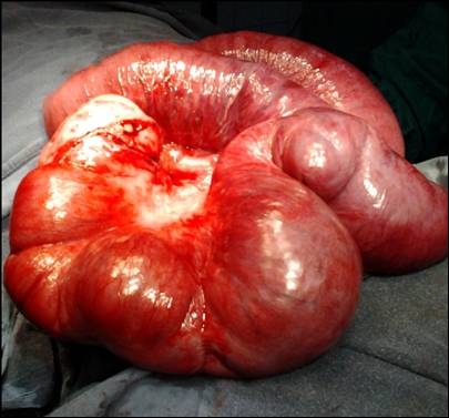
Figure
1: A feet long thick bowel mass
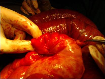
Figure
2: Unveiling of accordion like packed bowel within a pearly white membrane.
Discussion
Abdominal cocoon, also referred
to as sclerosing encapsulating peritonitis, is a rare cause of acute abdomen in
childhood, mostly involving young adolescent females. There is an encasement of
small bowel (sometimes whole of the abdominal viscera) by a fibrocollagenous
cocoon like sac which is usually formed by a nonspecific chronic inflammatory
reaction. It can be idiopathic or secondary to practolol intake, chronic
peritoneal dialysis, ventriculoperitoneal and peritoneovenous shunts,
sarcoidosis, liver cirrhosis, leiomyomata of uterus,
endometriotic cyst, tumors of ovary, abdominal tuberculosis [1,2]. In our case the cocoon was probably idiopathic in
absence of features specific to other disease processes.
Abdominal cocoon usually
present as sub-acute intestinal obstruction. However presentation could be chronic
constipation, anorexia, weight loss and abdominal mass in rare instances. Devay et al in 2006 could find only 47 reported cases of
abdominal cocoon. Out of these cases only few presented with mass in right
iliac fossa [2]. In our case patient presented with pain in right iliac region
and vomiting thus simulated acute appendicitis. Due to the position of the
cocoon at right iliac fossa and some ongoing inflammatory process causing
peritoneal irritation, the patient had sudden attack of pain in that region.
References
1.
Serafimidis C, Katsarolis I, Vernadakis S,
Rallis G, Giannopoulos G, Legakis N, et al. Idiopathic Sclerosing
encapsulating peritonitis (or abdominal cocoon). BMC Surg 2006, 6: 3.