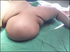
Figure 1: A large lumbo-sacral skin covered mass.
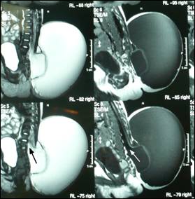
Figure 2: MRI sagittal section showing two cysts. The inner cyst
was in continuation (arrow) with the central canal of the spinal cord.
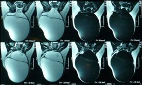
Figure 3: MRI in coronal section showing double compartment swelling.
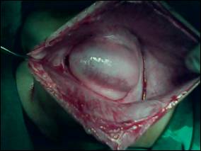
Figure
4: Another cyst inside the major cyst- the terminal myelocystocele.