Epigastric Heteropagus
Twin
Authors: Muhammad Qasim,* Mahmood shaukat
APSP Journal of Case Reports 2011;
Volume 2 (3): 24
Address: Department of Paediatric Surgery, Mayo Hospital Lahore,
Pakistan
Email:* qasim_179@yahoo.com
Date
of Submission: 26-9-11
Date
of Acceptance: 26-10-11
Citation: Qasim M, Shaukat M. Epigastric heteropagus twin. APSP J Case Rep 2011;2:24
Abstract
Parasitic
twining is a rare type of monozygotic monochorionic monoamniotic asymmetrical conjoined
twin. We report a case of epigastric heteropagus twin. An ultrasound scan showed
a defect of 1.5 cm in the epigastrium. CT showed soft tissue lobulated mass
with fat and air components coming out of the epigastric defect. At operation
rudimentary alimentary canal with no viscera, was found in the parasite. The
parasite was easily separated from the host.
Key
words: Conjoined twin, Monochorionic monoamniotic, Epigastric heteropagus
twin.
Introduction
Conjoined
twins have expected frequency of 1 in 50000 to 100000 live births. Potter and
Craig used the term of heteropagus for asymmetrical conjoined twins. Parasitic
twins account for 1-2% of all conjoined twins. The dependent undeveloped twin, the
parasite, is attached to independent developed twin called autosite
at different sites. Parasite attached to host’s epigastrium is rare and called
epigastric heteropagus [1,2]. We are reporting a case
of epigastric heteropagus twin to share the surgical findings.
Case
report
A full
term male baby was born with an undeveloped parasite attached to epigastric
region. He was brought to our hospital at the age of one month. The child was
healthy weighing about 5 kg. The parents and other siblings were healthy.
On
examination there was a 6x4 cm mass attached to the abdomen having rudimentary
upper limbs, lower limbs, head and external genitalia (Fig.1). An ultrasound
scan showed a defect of 1.5 cm in the epigastrium through which the gut was
herniating. CT scan showed a soft tissue lobulated mass with fat and air
components coming out of the epigastric defect. There were no calcifications.
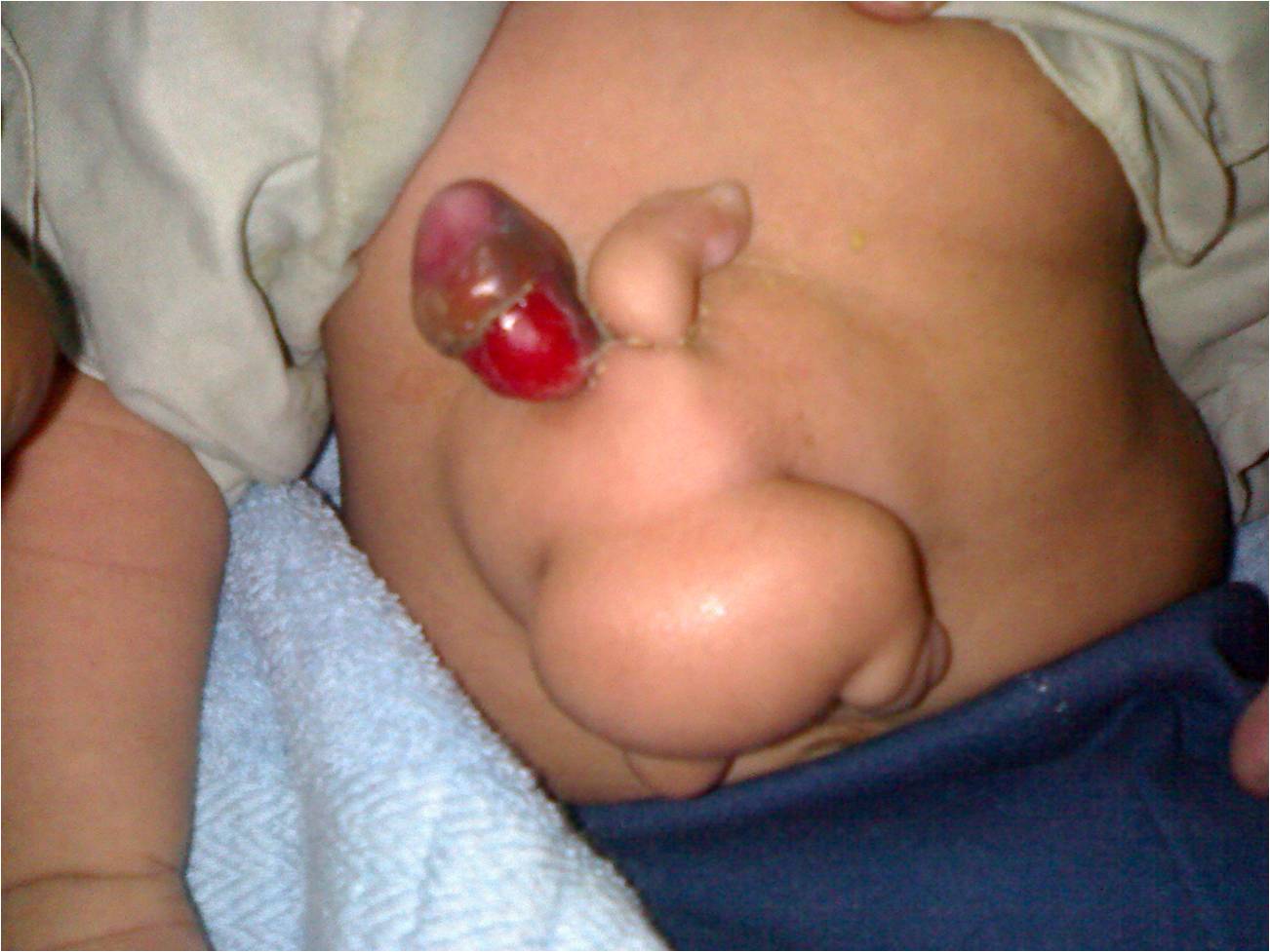
Figure 1: Parasite
showing rudimentary limbs and phallus
Separation
of the parasite from the host was done easily. The parasite had soft tissue
mass with no fully developed viscera. Rudimentary alimentary canal was present
in the parasite (Fig. 2). There were no sharing of viscera between host and
parasite. The postoperative recovery was uneventful and the baby was discharged
on 8th postoperative day.
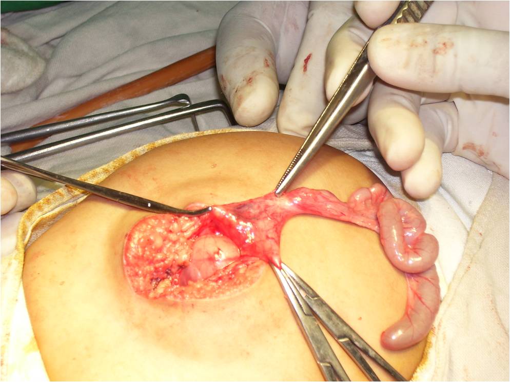
Figure 2:
Rudimentary bowel of parasite
Discussion
Conjoined twins are
monozygotic twins in which the inner cell mass does not completely split. The
two embryos are joined by a tissue bridge. Incomplete division of embryonic
disk after 13th day post conception results in the formation of
conjoined twins. Spencer proposed an alternative theory of fusion of two
originally separate monozygotic embryonic disks, to explain the conjoined twin
etiology. Some authors suggested that parasitic twin occur as a result of
selective ischemic damage in-utero leading to death or partial resorption of,
one of the twins, resulting in an incomplete parasitic twin attached to a fully
developed twin [3-5].
Conjoined
twin can be symmetrical or asymmetrical. Asymmetrical conjoined twins are
called parasitic or heteropagus twins. It is further classified as
1- Externally attached parasitic twin
2- An enclosed fetus in fetu
3- An internal teratoma
4- Ancardiac connected via the placenta
The
site and extent of twin fusion is extremely variable and the nomenclature is
usually based on fused anatomical region as in this case the parasite was
attached to the host in epigastric region so named as epigastric heteropagus [6,7]. In our case, parasitic twin had rudimentary limbs and
external genitalia. As in many of the reported cases, parasitic twin had limbs
and trunk formed to variable extent but was acephalic
and acardiac. In our case the blood supply of the
parasite was from falciform ligament as noted in most
reported cases [8,9]. Epigastric heteropagus is a rare
congenital malformation. The outcome and prognosis depends on the extent of
visceral sharing and associated anomalies.
References
1. Potter EL, Craig JM, Editors. Pathology of
Fetus and Newborn. 3rd ed. Chicago:Year Book;1975. p. 220-37.
2. Gupta DK, Lal A, Bajpai M. Epigastric heteropagus twins - a report of four
cases. Pediatr Surg Int 2001;17:481.
3. Zimmerman AA. Embryologic and anatomic
considerations of conjoined twins. National Foundation 1967;3:18.
4. Spencer R.
Parasitic conjoined twins: external, internal (fetuses in fetu
and teratomas), and detached (acardiacs).
Clin Anat 2001;14:428–44.
5. Spencer R. Theoretical and analytical
embryology of conjoined twins. Clin Anat 2000;13:36.
6. Satter E, Tomita S. A case report of omphalopagus heteropagus (parasitic) twin. J Pediatr Surg 2008;43:E37-9.
7. Bega G, Wapner R,
Lev-Toaff A, Kulhman K.
Diagnosis of conjoined twins at 10 weeks using three dimensional
ultrasound: a case report. Ultrasound Obstet Gynecol 2000;16:388.
8.
Cury EK, Schraibman V. Epigastric heteropagus twinning. J Pediatr Surg 2001;36:11.
9.
Jan IA, Haq A, Sharif A,
Khan S, Khan O. Epigastric
heteropagus parasitic twins- A report of three cases. J Pediatr Surg Spec
2008;2:28-31.