Figure
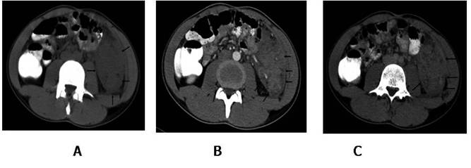
1: Axial computed tomography image show a soft tissue mass, located between
left psoas muscle and lateral abdominal wall. The lesion partially surrounds
the sigmoid colon (A). Contrast enhanced CT arterial (B) and venous (C) phase
show marked heterogeneous involvement.
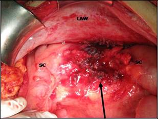
Figure 2: Arrow indicates the location of removed mass. It
was over sigmoid colon (SC) and lateral abdominal wall (LAW).
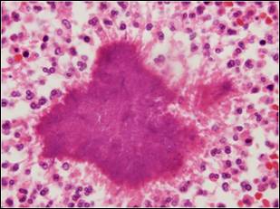
Figure 3a: A colony of
actinomyces and mixed inflammatory response at the periphery which include
histiyocytes, lymphocytes, plasmocytes, eosinophils and neutrophils. ( H&E,
original magnification x100 )
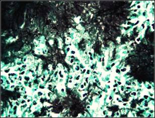
Figure 3b: Colony of filamentous
microorganisms stain black with Grocott’s methenamin silver conventional staining
method ( magnification x100 immertion ).