Ewing’s Sarcoma in Scapular
Region
Authors: S H Waqar,* M A Zahid
APSP Journal of Case Reports 2011;
Volume 2 (3); 22
Address: Department of General Surgery, Pakistan Institute of
Medical Sciences Islamabad, Pakistan
Email:* drshwaqar@gmail.com
Date of Submission: 12-08-11
Date of Acceptance: 14-10-11
Citation: Waqar SH, Zahid
MA. Ewing’s sarcoma in scapular region. APSP J Case
Rep 2011;2:22
Abstract
Ewing's
sarcoma (ES) primarily affects bones and commonly presents in adolescents and
young adults. This paper reports a rare
case of extra osseous ES of the scapular region in a 9 years old girl. She was
treated by a multidisciplinary approach including surgery, chemotherapy and
radiotherapy. She was followed up for two years and remained well.
Key
words: Ewing’s sarcoma, Extra-osseous, Chemotherapy.
Introduction
ES
is a rare and highly malignant small round cell tumor that primarily affects
the skeletal system. In primary extra osseous ESs of soft tissue underlying
bone involvement is not found. James Ewing described it in 1921 as a
tumor arising from undifferentiated osseous mesenchymal
cells; however, recent studies suggest that Ewing’s tumor may be of neuroectodermal origin being derived from the primitive
neural tissue. It accounts for 4 to10% of all types of bone cancers, with long
bones and pelvis being the most common sites. It is the second most common bone
tumour of childhood and adolescence, with male
preponderance of 1.6:1. It is rarely seen before the age of 5 years and after the
age of 30 years [1-4].
The
ES usually arises in the metaphysis or diaphysis of long bones of extremities.
The lungs, bones and bone marrow are the most common sites of metastasis. An
extensive review of the literature showed only few reported cases of the extra-osseous
ES in patients under the age of 10 years. This report describes a case of extra
osseous ES of the scapular region in a 9 years old girl. This case elucidates
the importance of professional knowledge of the relevant aspects of ES.
Case
Report
A
9 year old girl presented in the surgical outpatient department with history of
a progressive swelling over right scapular region for the last three years.
Swelling started as a small lump that increased in size during last six months.
Swelling was not associated with fever, malaise and fatigue. There was no
history of exposure to any carcinogenic agent or radiation.
Past history was not significant. It was her school teacher who asked her
parents to seek medical advise
for the swelling, as she was facing difficulty in writing.
The
general examination of the child was normal. Local examination revealed a
globular non tender swelling over the right scapular region, measuring 40x36x38
cm, having firm to hard consistency. It was mobile with well defined margins
and not attached to deeper structures. Overlying skin was mobile, shiny with
multiple visible vessels and a small ulcer noted in the center of the swelling (Fig.
1). There was no neurovascular deficit distal to the tumour.
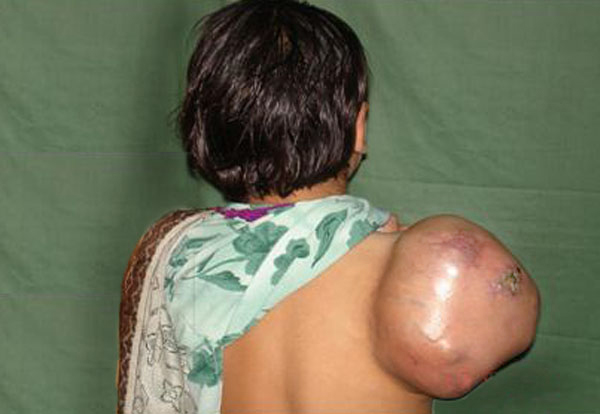
Figure 1:
Mass at Scapular Region
Blood
complete picture revealed mild anemia. Other blood tests including renal
function test, liver function test, and serum calcium and serum alkaline
phosphatase were within normal limits. X-ray and CT scan of the scapular region
showed soft tissue swelling without any bony involvement (Fig. 2,3). Fine needle aspiration suggested malignant soft tissue tumour. Incisional biopsy confirmed the diagnosis of ES. The
lesion was excised and residual defect was left as such with a plan skin
grafting at a later stage. The recovery was smooth and patient was discharged
on fifth postoperative day. Histopathology confirmed the diagnosis of ES. All
resection margins were free of tumour. Immunohistochemistry
was not done. She did not report back for follow up. After three months of
surgery she attended the surgical outpatient department with almost healed wound
(Fig. 4). She was referred to oncologist for chemotherapy. She received VAC
regimen (vincristine, adriamycin and cyclophosphamide),
repeated every 3 weeks for 6 cycles and local radiotherapy was also given to
the excised area. The patient responded well to treatment and has not shown any
recurrence after two years of follow up.
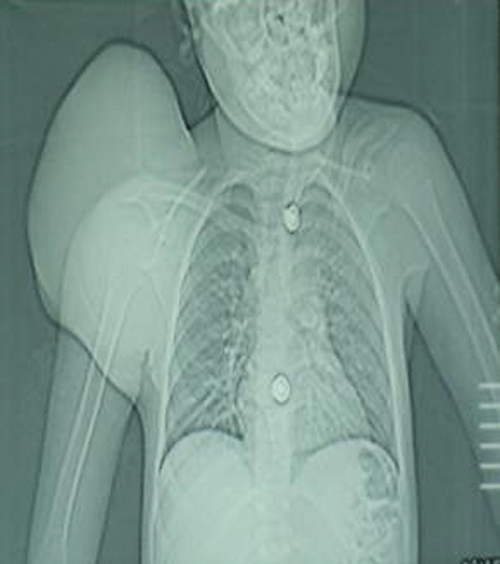
Figure 2:
CT scout view showing soft tissue shadow
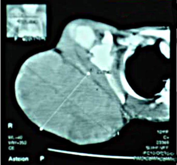
Figure 3:
CT scan showing soft tissue tumour
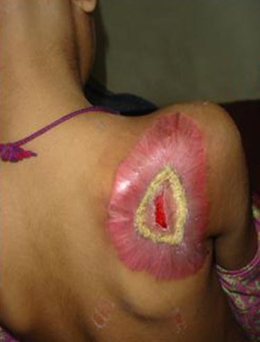
Figure 4:
Three months postoperatively
Discussion
Soft
tissue ES is a rapidly growing, round-cell, malignant tumour
which can reach 10 cm size by the time diagnosis is
made. Commonly affected extra-osseous sites are the paravertebral
spaces, lower extremities, head and neck, and pelvis. One paper reported an ES
involving the scapular region on literature serach.
The most frequent presenting symptom is a rapidly growing mass with local pain.
ES may also present with systemic signs and symptoms such as weight loss and
fever [5-7]. The index case aged nine
year presented with a mass without any constitutional symptoms.
Extraskeletal
ES is confirmed by characteristic features on histological analysis, histochemistry, immunohistochemistry
and electron microscopy. Differential diagnoses include other small, blue round
cell tumours (SBRCTs) and other members of the Ewing
family of tumours such as the primitive neuroectodermal tumour (PNET). Findings
of X-ray and CT scan of our case showed the tumour as
soft tissue swelling over scapular region. Molecular and cytogenetic analysis
should be considered as the standard practice in the diagnostic evaluation of
ES [5,8].
The
mainstay treatment should include multi-agent chemotherapy and aggressive
surgical treatment. Tumours
that are not appropriate for surgical resection or have positive margins are
treated with radiation. The results of surgery alone for extra-osseous ES are
poor in most of the cases, while patients receiving multimodal chemotherapy and
radiotherapy have a much better prognosis. With the combination of local
surgical treatment and systemic chemotherapy, long-term survival has improved
from 10% to 50%-60% or greater. The prognosis for extra-osseous ES appears more
favourable than that of ES in bone. More recently, Ifosfamide has emerged as a an effective
chemotherapeutic agent, specially in patients resistant
to other drugs. Better long term survival can be achieved in patients
presenting with non metastatic disease [8-10]. Our patient responded well to
the treatment and remained well after two years of follow up.
In
summary, surgical resection, multi agent chemotherapy, and radio-therapy are
the mainstay of treatment of ES. The treatment plan should be individualized
for each patient, which should be based on age, location, stage, size of the
tumor and response to therapy.
Acknowledgements: We acknowledge the work of our hospital photographer and Dr Mukhtar (Postgraduate resident) for photography.
References
1.
Naru T, Nawaz FH, Rizvi J. Juvenile Ewing’s
sarcoma presenting as a pelvic mass. J Coll Physicians
Surg Pakistan 2007;17:53-4.
2.
Maheshwari V, Siddiqui F, Adreena, Sherwani RK, Jain A, Alam
K. Extraskeletal Ewing’s sarcoma- A case report. Internet J Orthoped Surg
2009;14:1.
3. Extraskeletal Ewing’s sarcoma/primitive neuroectodermal tumor family. In: Enzinger
and Weiss’s Soft Tissue Tumors, 4th ed. St Louis: Mosby 2001; 1289-91.
4.
Túllio BM, Vieira FA, Rogério de FP, Vitorino CS, Mota LA. Ewing's sarcoma of the mandible in a young child. Braz Dent J 2010;21:74-9.
5.
Askri A, Farhat LB, Ghariani B, Rabeh A, Dali N, Said
W, et al. Extraskeletal Ewing sarcoma of the abdominal wall. Cancer Imaging
2008;8:156-8.
6.
Cheung
CC, Kandel RA, Bell RS, Mathews RE, Ghazarian MD. Extraskeletal Ewing sarcoma in a 77-year-old
woman. Arch Pathol Lab Med 2001;125:1358-60.
7. Asif N, Khan AQ, Siddiqui
YS, Mustafa H. Metastasis
from scapular Ewing’s sarcoma presenting as sutural diastasis: An unusual presentation. Int
J Shoulder Surg 2010;4:18-21.
8.
Perouli E, Chrysikopoulos H, Vlachos
A, Koskinas A, Batistatou
A, Polyzoidis K. Imaging findings in paraspinal extra osseous Ewing sarcoma. JBR-BTR 2006;89:310-2.
9.
Aydinli B, Ozturk G, Ilhan Yildirgan M, et al.
Extraskeletal Ewing’s sarcoma in the abdominal wall: a case report. Acta Oncol 2006;45:484-6.
10.
Colovic RB, Grubor NM, Micev MT, Matic SV, Atkinson HDE,
Latincic SM. Perigastric extraskeletal
Ewing’s sarcoma: A case report. World
J Gastroenterol 2009;15:245-7.