Spontaneous Intravesical Knotting of
Urethral Catheter
Author: Yogesh Kumar Sarin
APSP Journal of Case Reports 2011; Volume 2 (3): 21
Address: Department
of Pediatric Surgery, Maulana Azad Medical College,
New Delhi-110002
Email address: sarinyk@yahoo.com
Date of Submission: 09-07-11
Date of Acceptance:
15-07-11
Citation: Sarin YK.
Spontaneous intravesical knotting of urethral catheter.
APSP J Case Rep 2011;2:21
Abstract
Infant feeding tubes (IFT) have been
universally used as urethral catheters in neonates and children for several
decades. Though generally a safe procedure, it may cause significant morbidity
if the catheter
spontaneously knots inside the bladder. We report this complication in three
children including a neonate.
Key
words: Urinary catheter;
Catheterization complication, Intravesical knotting, Posterior sagittal anorectoplasty.
Introduction
Catheters
inserted for various purposes, urological as well as non-urological, are known
to rarely knot spontaneously inside the human body with an estimated incidence of 0.2 per 100,000 catheterizations [1]. Raveenthiran could
find only 40 cases of knotted urinary catheters on a recent review of the world
literature [2]. The report intends to generate awareness of a potentially
preventable complication that can result in significant morbidity with a list
of recommendations to minimize this risk.
Case Report
Case 1: An eight-month-old male infant, a case of anorectal
agenesis with rectoprostatic urethral fistula with status sigmoid loop colostomy,
underwent posterior sagittal anorectoplasty. He was catheterized with a 6 Fr
infant feeding tube intra-operatively. The surgery and the post-operative
period were uneventful. Gentle traction on the catheter however failed to
retrieve the catheter on seventh post-operative day. On examining along the
urethra, the knotted catheter could be palpated at the perineum. Pelvic roentgenogram
confirmed the diagnosis of knotted catheter in the urethra. Several attempts at
forceful introduction of sterile saline and contrast material under fluoroscopy
failed to unwind the loop.
Under short dissociate anesthesia, another
attempt was made to untie the knot and straighten the catheter with angiography
wire through the catheter lumen. Failing this maneuver, the catheter was
divided flush with the glans penis and the knotted
catheter was gently manipulated out through a small perineal urethrostomy (Fig.1).
A percutaneous suprapubic
tube was inserted and was left in place for a week. The child had been passing
urine in good stream on follow up.
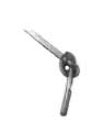
Figure 1: Knotted catheter after retrieval through
perineal urethrostomy
Case 2: A male newborn weighing 1.8 kg was operated for ileal
atresia on day-1 of life; resection of atretic segment and
end-to-back ileo-ascending colic anastomosis was
done. The patient was re-operated after two weeks for anastomotic leak. Three
days later, an
attempt to remove the catheter (6 Fr IFT) was met with resistance. From the
past clinical experience, catheter knotting was suspected. On this occasion, manipulation with angiography wire
through the catheter lumen succeeded and the catheter removed. The child was discharged
after a month of admission. Unfortunately, a week later he was brought moribund
to the casualty where he succumbed to severe dehydration and refractory shock.
Case 3: One and half year old boy underwent endoscopic valve
incision for posterior urethra valves. The child was lost to follow-up for 5
years when he presented again with poor urinary stream. He could not be
catheterized and was diagnosed to have urethral stricture at bulbo-membranous
junction on retrograde urethrography. Endoscopic incision of hypertrophied
bladder neck and visual internal urethrotomy of
stricture was done; there were no residual posterior urethral valves. Three days
later, an
attempt to remove the catheter was met with resistance. The catheter was
removed using local and systemic analgesia and gentle steady traction. The tip
of the catheter was found knotted. The patient voided clear urine spontaneously
and comfortably after few hours. He later underwent endoscopic management of
bilateral major grades of vesico-ureteral reflux (deflux injection). He is
under close follow up.
Discussion
Intravesical knotting of catheters have been
reported more commonly in males than females, and more commonly in neonates and
children than adults [2]. Intravesical knotting has been reported not only in catheters left
for bladder drainage, but also after brief maneuvers such as clean intermittent
catheterization, and cystourethrography [3-5].
This is the first instance
that this complication has been encountered following posterior sagittal
anorectoplasty.
Although
knotting of urethral catheters is rare, removal represents significant
morbidity, such as general anesthesia, radiation exposure during fluoroscopy,
and transient hematuria [1]. Potential for further
complications such as stricture formation also needs to be considered. Knotted
urinary catheters may also jeopardize delicate surgical reconstructions [3, 6].
Unfortunately, many doctor colleagues and nursing staff are unaware of this
problem or its proper management. A telephone survey of 24 tertiary- care
Emergency Departments in Canada revealed that none of them were aware of
catheter knotting and 22 had no protocol established for safe catheterization [1].
Several
hypothetical explanations have been offered for the knotting of catheters. The tendency of a catheter to knot probably depends on
its flexibility, smaller diameter and redundancy within the bladder. The
probable mechanism involves an extra length of catheter coiling around itself
and then the catheter end looping through these coils [4]. The coils tighten
cinching down in a knot when counter traction is applied to remove the
catheter. If the diameter of this knot exceeds that of urethra the catheter
gets stuck. Bladder spasm has been also been attributed as a risk factor [7]. Water-current
generated by the flow of urine around the catheter may also play a role in the genesis
of catheter knotting [2]. Raveenthiran suggested that the catheters slender
than 10 Fr, over-distended bladder and insertion of excessive length (greater
than 10 cm beyond bladder neck) of catheters must be considered as risk factors
for catheter knotting [2].
Several techniques
have been described to retrieve the knotted catheter. They include sustained
traction under anesthesia, unraveling the knot using a guide-wire through the
catheter under fluoroscopy, endoscopic retrieval and suprapubic
cystotomy [4,7-9]. Guide-wire manipulation is useful
only at the early ‘open-loop stage’ of knot formation when the knot is not
tight enough [8] and succeeded in one of our cases. Sustained traction also
worked once, but such a manipulation with or without urethral dilatation carries
the risk of urethral damage. Moreover, this technique is not useful when the
knot is bulky or when two catheters knot together [2]. Suprapubic
cystotomy has been known as a simple, safe and cost-effective method of
retrieving knotted bladder catheters [2], though in the modern era, this could
be replaced by vesicoscopy.
The attention should be directed towards
prevention of this complication by careful selection of the catheters and
gaining better understanding of urethral anatomy and safe insertion lengths. The
insertion lengths of 6 cm in a male newborn and 5 cm in a female newborn have
been recommended [10]. In extremely premature babies with birth weight of
<750 grams the insertion length of <2.5 cm in girls and <5 cm in boys
is recommended [10]. It is also equally important to secure the catheter well
in order to prevent inadvertent advancement of the catheter into the bladder [1].
References
1.
Arena
B, McGillivary D, Dougherty G. Urethral catheter
knotting: be aware and minimize risk. Can J Emerg Med
2002;4:1-5.
2.
Raveenthiran V. Spontaneous knotting of
urinary catheters: clinical and experimental observations. Urol
Int 2006;77:317-21.
3.
Ball
RA, Horton CE Jr, Mandell
JA. Transurethral removal of knotted bladder drainage catheter in a male
following bladder neck reconstruction. Urology 1993;41:234-6.
4.
Klein EA, Wood DP, Kay R. Retained straight
catheter complication of clean intermittent catheterization. J Urol 1986;135:780-1.
5.
Cass A, Vitko R.
An unusual complication of cystourethrography. Minn
Med 1972;55:355-6.
6.
Singh
RB, Pavithran NM, Parameswaran
RM. Knotting of feeding tube used for bladder drainage in hypospadias
repair. J Indian Assoc Pediatr Surg
2005;10:199.
7.
Arda IS, Ozyaylali I.
An unusual complication of suprapubic catheterization
with Cystofix: catheter knotting within the
bladder. Int J Urol 2001;8:188-9.
8.
Harris VJ, Ramiro J. Wire manipulation of
knot in a catheter used for cystourethrography. J Urol
1976;116:529.
9.
Dogra PN, Nabi G, Goel R. Endoscopic removal of knotted urethral catheter: a
point of technique. Urol
Int 2003;71:8-9.
10.
Carlson D, Mowery B. Standards to prevent
complications of urinary catheterization in children: should and should-knots.
J Soc Pediatr Nurs 1997;2:37-41.