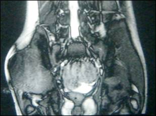
Figure 1: MRI of right gluteal region.
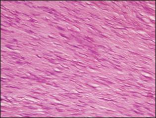
Figure 2: Well differentiated and poorly cellular fibrous tissue with no increased mitotic activity, nuclear pleomorphism or necrosis. (H& E x 40)