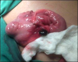
Figure
1: An infarcted lymph node just adjacent to cecum.
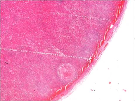
Figure 2: Showing a thin rim of
viable subcapsular lymphoid tissue with hemorrhage in the marginal sinus (x40).
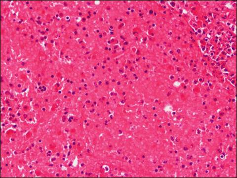
Figure 3: Showing extensive
necrosis of medullary and cortical lymphoid cells with extravasated red blood
cells (x 400).