
Figure 1: CT scan showing solid and
cystic components with internal calcifications.

Figure 2: Mass arising from the
posterior gastric wall.
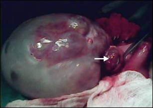
Figure 3: The
posterior gastric wall was opened to show endogastric component (arrow).
Figure 1: CT scan showing solid and
cystic components with internal calcifications.
Figure 2: Mass arising from the
posterior gastric wall.
Figure 3: The
posterior gastric wall was opened to show endogastric component (arrow).