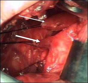
Image 1: Intra-operative picture
showing the proximal esophageal pouch (thin white arrow) and blind tracheal
diverticulum (thick white arrow).
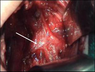
Image 2:
Intra-operative picture showing right TB (white arrow).
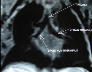
Image 3:
Post-operative MRI showing right upper lobe bronchus originating at the level
of the carina.
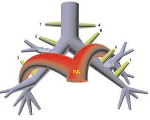
Image 4: Schematic
representation of aberrant bronchi to the upper lobes: prearterial (true
right tracheal) (1), preeparterial (right “tracheal”) (2), posteparterial (3),
eparterial (true left tracheal) (4), eparterial (left “tracheal”) (5),
prehyparterial (6), and posthyparterial (7) bronchi.
LULB = left
upper lobe bronchus, PA = pulmonary artery, RULB = right upper lobe bronchus.
(reproduced
with permission of publishers of Radiographics).