
Image 1: Clinical photograph showing giant occipital
encephalocele.
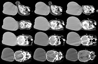
Image 2: CT scan brain showing large encephalocele sac
with protrusion of the contents.
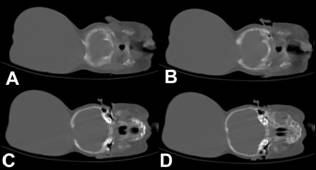
Image
3: CT scan brain (bone window) showing large defect in the occipital bone with
sclerosed margins.
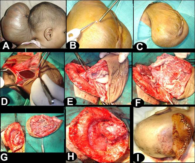
Image
4: Intra-operative photographs showing operative steps (B) small amount of the
CSF was let out through a small opening in the sac, (C) note the reduction in
the size of swelling, (D) sac was opened, (E) base of sac was defined, note the
presence of large vessels near to the opening in the skull, (F and G) redundant
brain tissue was excised while preserving the large vessels, (H and I) dura and
skin were closed.